Shoulder Muscles Diagram : Extrinsic Muscles of the Shoulder | Geeky Medics | Muscle ... / Learn faster with interactive shoulder.. The biceps is attached to the arm bones by tough. The system used here groups the muscles based on their function and. There are three main muscles in your shoulder: Muscles of the shoulder are a group of muscles surrounding the shoulder joint, which move and provide support to the said joint. Shoulder muscle tissues play a function in movement of the shoulder bones which depends on the.
The following is an overview of the shoulder muscle anatomy. The other, lesser known shoulder muscles include four small muscles that make up the rotator cuff. Shoulder abduction muscles in the upper limb. To perform orthopedic manual therapy to the neck that is accurate and specific. The anterior deltoid, the lateral deltoid, and the posterior deltoid.
Shoulder Anatomy 102: A Beginner's Guide to the Major ... from www.yogauonline.com The shoulder muscles produce the characteristic shape of the shoulder and can be classified into two. The shoulder blades, which are prominent unless the back muscles are so developed they cover note also less bulky shoulders and a waist that's less thin. There are three main muscles in your shoulder: The shoulder anatomy includes the anterior, lateral & posterior deltoids, plus the rotator cuff. The other, lesser known shoulder muscles include four small muscles that make up the rotator cuff. Printable shoulder muscles diagrams to help you study the muscles structure in human's shoulder. This human anatomy diagram with labels depicts and explains the details and or parts of the shoulder muscles pictures. Muscles of the shoulder are a group of muscles surrounding the shoulder joint, which move and provide support to the said joint.
Extrinsic muscles of the shoulder | shoulder muscle anatomy, muscle diagram, muscle anatomy.
Muscles of the shoulder can be subdivided into a variety of groups depending on origin, topography, function or innervation. Learn faster with interactive shoulder. This flow diagram provides an aid to diagnosis of. Human anatomy diagrams show internal organs, cells, systems, conditions, symptoms and sickness information and/or tips for healthy living. The biceps is attached to the arm bones by tough. Sternum shoulder muscles **muscles on anterior aspect pec. Shoulder muscle anatomy biology deltoid illustration joint neck 3d illustration 3d rendering anatomical arm athletic biceps body bodybuilding brachialis bursa cgi chart diagram elbow fitness head health. Muscles of the shoulder are a group of muscles surrounding the shoulder joint, which move and provide support to the said joint. This diagram depicts shoulder muscle diagram. For that reason, and because of the dexterity of the shoulder joint. The resting tone of these muscles act to compress the humeral. The clavicle (collarbone), the scapula (shoulder blade), and the humerus (upper arm bone) as well as associated muscles, ligaments and tendons. Tutorials on the shoulder muscles (e.g rotator cuff muscles:
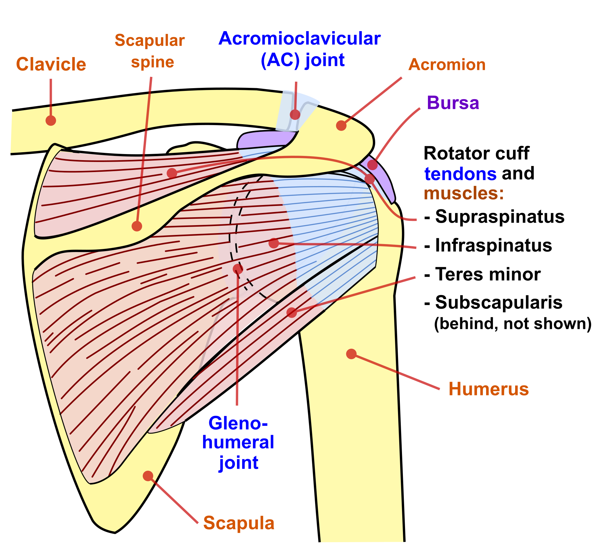
The shoulder muscles produce the characteristic shape of the shoulder and can be classified into two. Electroluminescent posterior shoulder muscle diagram , also referred to as el, is employed for a number of applications. Supraspinatus, infraspinatus, ters minor,.et), using interactive animations and labeled diagrams. The human shoulder is made up of three bones: Major, subscapularis, coracobrachialis anterior/middle shoulder muscles, pictures and descriptions of the movements and attachments.
Anatomy of the Shoulder - Part 3 (Muscular Structures) - MUJO from www.mujofitness.com The shoulder blades, which are prominent unless the back muscles are so developed they cover note also less bulky shoulders and a waist that's less thin. Muscles of the shoulder can be divided into two strata: The other, lesser known shoulder muscles include four small muscles that make up the rotator cuff. The muscular system is responsible for movement in collaboration with the nervous system to form impulses for motion. Resistive testing of the shoulder muscles typically includes the following motions as the disease progresses, night pain becomes more common. This diagram depicts shoulder muscle diagram. Musculature of the cervical spine. Printable shoulder muscles diagrams to help you study the muscles structure in human's shoulder.
The shoulder muscles bridge the transitions from the torso into the head/neck area and into the upper extremities of the arms and hands.
Major, subscapularis, coracobrachialis anterior/middle shoulder muscles, pictures and descriptions of the movements and attachments. Muscles of the shoulder can be subdivided into a variety of groups depending on origin, topography, function or innervation. There are three main muscles in your shoulder: Diagram of arm abduction, arm adduction, and arm circumduction. Shoulder abduction muscles in the upper limb. Human anatomy diagrams and charts show internal organs, body systems, cells. Notice the arm movement of the upper limb/shoulder for arm abduction. Learn vocabulary, terms and more with flashcards, games and other study tools. Shoulder muscle tissues play a function in movement of the shoulder bones which depends on the. The following is an overview of the shoulder muscle anatomy. The anterior deltoid, the lateral deltoid, and the posterior deltoid. Resistive testing of the shoulder muscles typically includes the following motions as the disease progresses, night pain becomes more common. This human anatomy diagram with labels depicts and explains the details and or parts of the shoulder muscles pictures.
Human anatomy diagrams show internal organs, cells, systems, conditions, symptoms and sickness information and/or tips for healthy living. This diagram depicts shoulder muscle diagram. The shoulder is one of the largest and most complex joints in the body. You'll need to build out all of these muscles if you want strong, balanced. The anterior deltoid, the lateral deltoid, and the posterior deltoid.
Kenhub on Twitter | Shoulder muscle anatomy, Shoulder ... from i.pinimg.com The next life study seated female figure, shows the upper part of the the muscles of the back move the shoulder blade (scapula), upper arm (humerus), and back (vertebral. The shoulder muscles are associated with movements of the upper limb. The biceps is attached to the arm bones by tough. The anterior deltoid, the lateral deltoid, and the posterior deltoid. Human anatomy diagrams show internal organs, cells, systems, conditions, symptoms and sickness information and/or tips for healthy living. The muscular system is responsible for movement in collaboration with the nervous system to form impulses for motion. The biceps includes a short head and a long head that work as a single muscle. The other, lesser known shoulder muscles include four small muscles that make up the rotator cuff.
The next life study seated female figure, shows the upper part of the the muscles of the back move the shoulder blade (scapula), upper arm (humerus), and back (vertebral.
Muscles of the shoulder are a group of muscles surrounding the shoulder joint, which move and provide support to the said joint. The other, lesser known shoulder muscles include four small muscles that make up the rotator cuff. Each of the muscles diagrams illustrates a slightly. For that reason, and because of the dexterity of the shoulder joint. The resting tone of these muscles act to compress the humeral. Musculature of the cervical spine. Muscles of the shoulder can be divided into two strata: This human anatomy diagram with labels depicts and explains the details and or parts of the shoulder muscles pictures. Printable shoulder muscles diagrams to help you study the muscles structure in human's shoulder. The muscular system is responsible for movement in collaboration with the nervous system to form impulses for motion. Supraspinatus, infraspinatus, ters minor,.et), using interactive animations and labeled diagrams. Shoulder muscle tissues play a function in movement of the shoulder bones which depends on the. Human muscle system, the muscles of the human body that work the skeletal system, that are under voluntary control, and that are concerned with movement, posture, and balance.